Advanced cell-based systems for reliable research
In vitro models

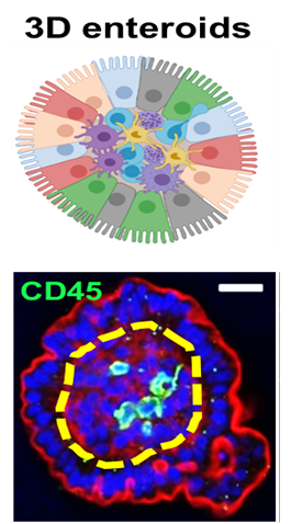
Chicken 3D intestinal organoids
Our chicken 3D intestinal organoids are developed following the validated method of Nash et al. (2021, 2023). These organoids originate from intestinal villi isolated from 18-day-old embryos and are cultured without an extracellular matrix such as Matrigel. This results in:
- Multiple villus-like budding structures
- Inside-out orientation for direct compound/microbial exposure
- Intact epithelial barrier function with immune-epithelial interaction
The inner core of the 3D organoids replicates the lamina propria, containing immune cells (e.g., natural killer cells, T cells, macrophages). This makes them an ideal model for studying innate immune responses to enteric microbes and compounds.
Applications of 3D intestinal organoids
- Assessing gut compatibility and efficacy of food additives
- Studying digestion and absorption of enzymes
- Evaluating immune responses to vaccines
- Testing pharmacokinetics and bioavailability of therapeutics
- Investigating gene expression and gut health
(Illustration: doi: 10.1038/s42003-021-01901-z)
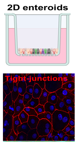
Chicken 2D intestinal organoids
Our chicken 2D intestinal organoid model, based on Orr et al. (2021), is derived from intestinal villi of 18-day-old embryos. These cells are seeded on plates or transwell inserts, creating a more physiologically relevant system compared to standard cell lines. This 2D system allows for:
- Conducting precise translocation studies of molecules, including feed compounds
- Monitoring intestinal barrier integrity in real-time
- Measuring Trans Epithelial Electrical Resistance (TEER) with precision
Applications of 2D Intestinal Organoids
- Assessing transwell barrier integrity
- Studying intestinal absorption
- Analyzing inflammatory responses
- Evaluating passive & active transport mechanisms
- Investigating infection and immune responses
(Illustration: doi: 10.1186/s13567-021-01010-z)
Our expert

Prof. dr. ir. Lonneke Vervelde
Senior scientist Poultry Health and R&D
Read the latest paper by Lonneke Vervelde about a new Enterococcus cecorum infection model at GD using chicken intestinal organoids: Invasion of Chicken Intestinal Cells Is Higher for Enterococcus cecorum Lesion Strains Compared to Cloacal Strains in an Organoid Model. Microorganisms2025, 13, 50